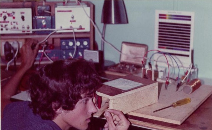
 Spark spectroscopy won me first place in a Science Fair event about 40 years ago. I just did it for the pretty colours.
“Continue reading” for more details about interpreting spectra to analyse gases and metals.
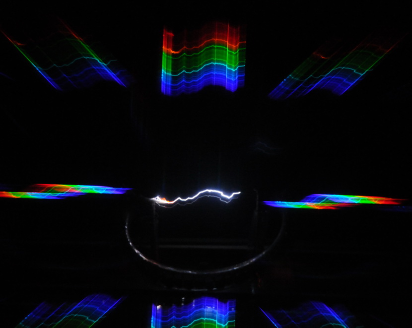
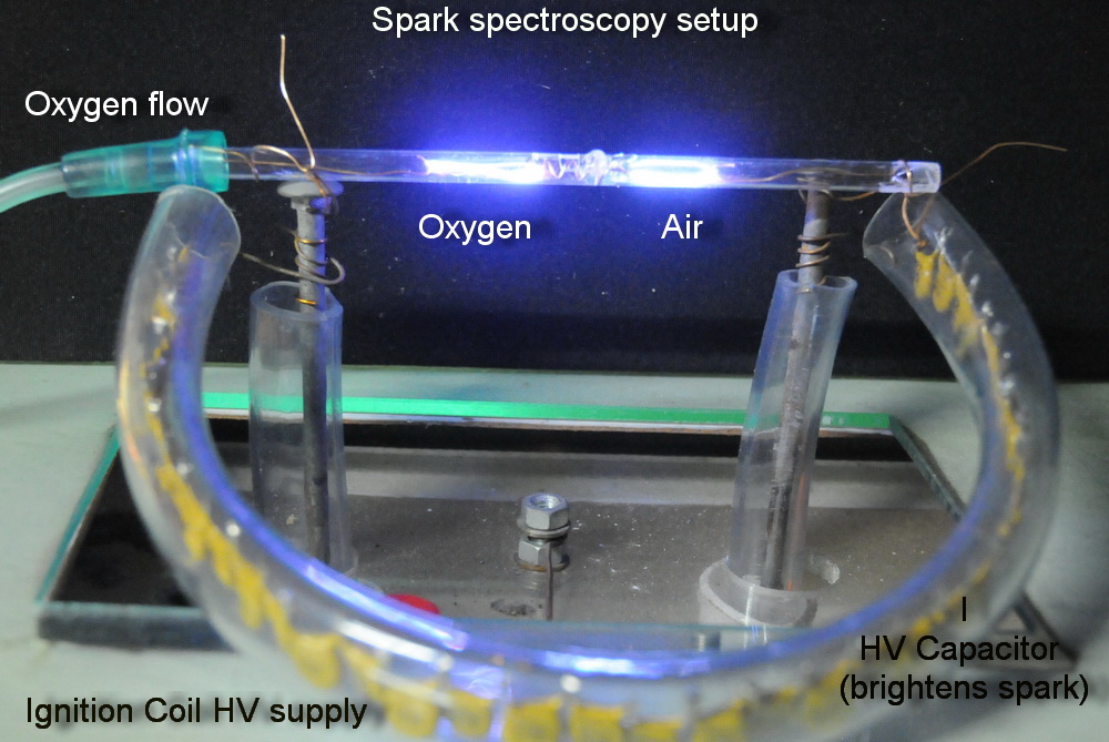
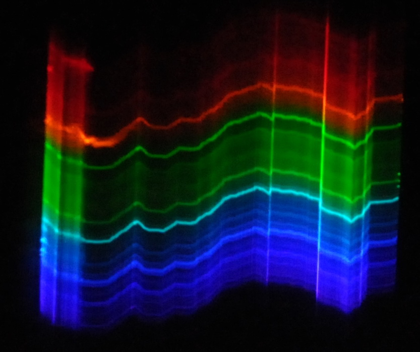
Take a photo of a transient bright spark about 2 inches long through a transmission diffraction grating and this is what you get. The multiple bands of parallel spark impressions are due to ionised oxygen and nitrogen. You can tell which is which by repeating this in pure oxygen.
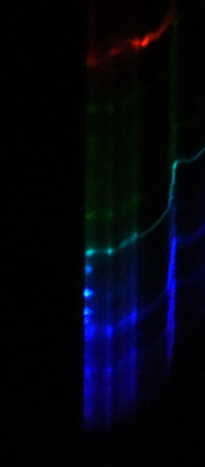

The interesting bit is the green dots at the extreme right. These are ionised copper from the spark contacting copper wire.
On the left side the spark contacts a galvanised bolt and the zinc coating is shown as blue dots at the left edge.
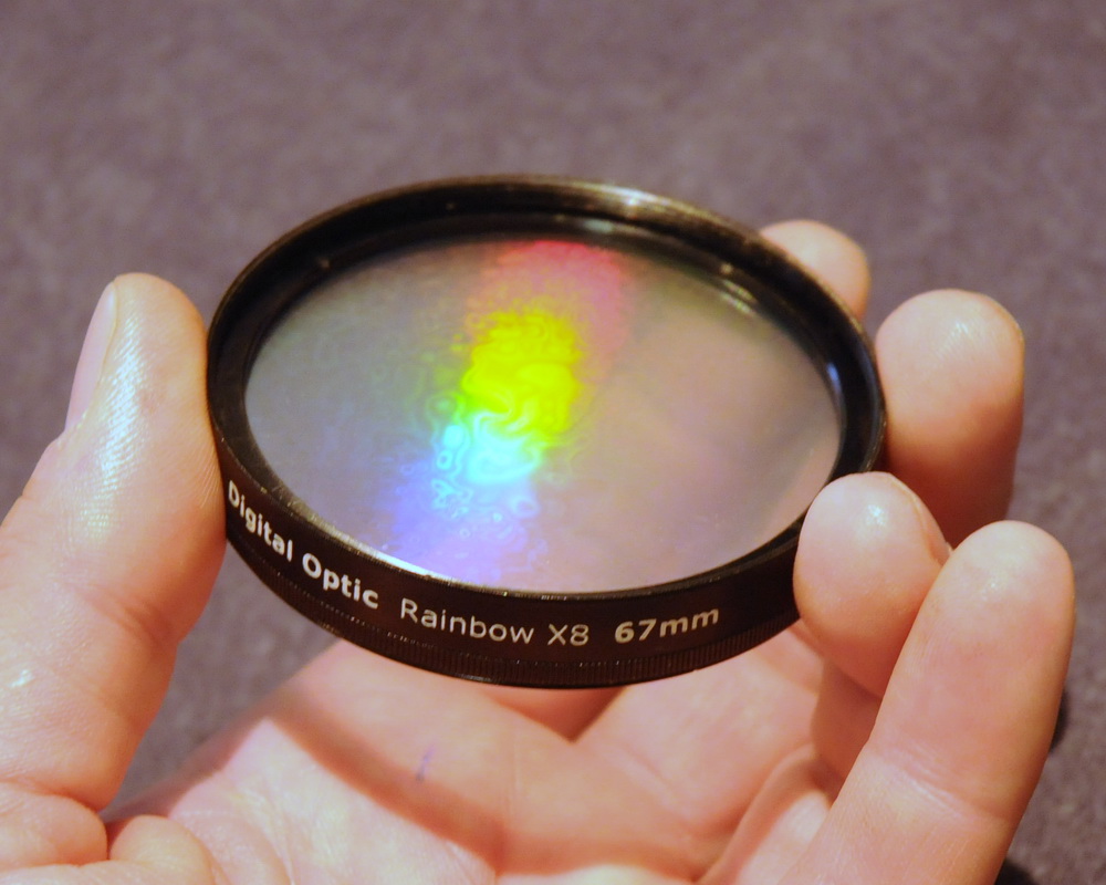
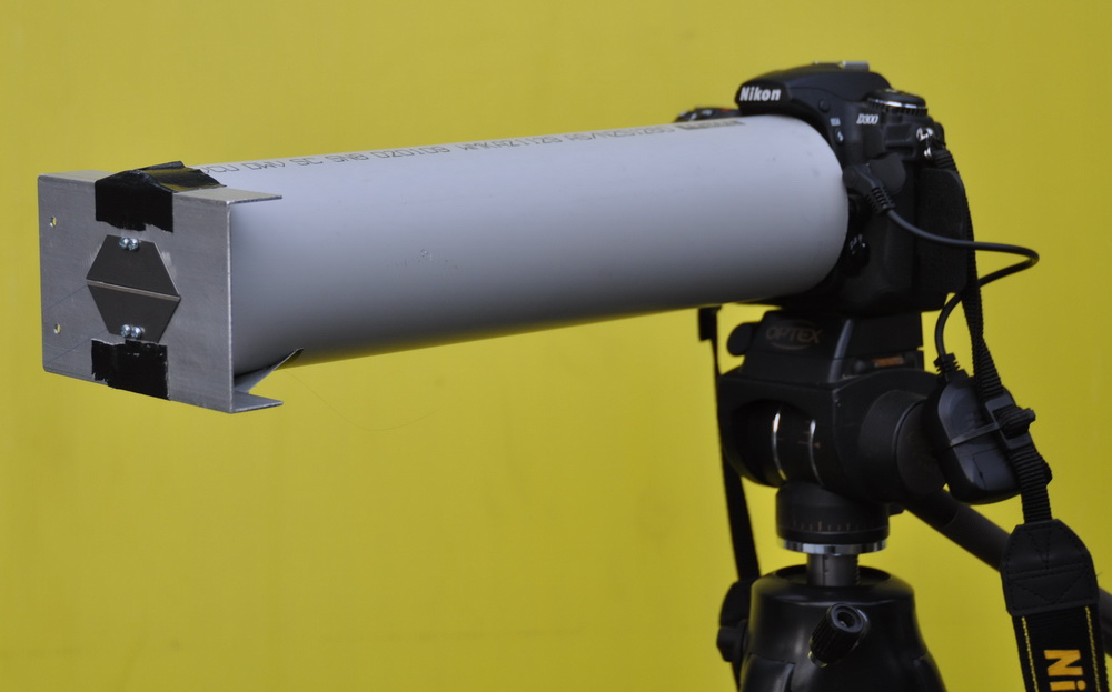
This is the full view of the multiple spectra from a special effects lens filter.
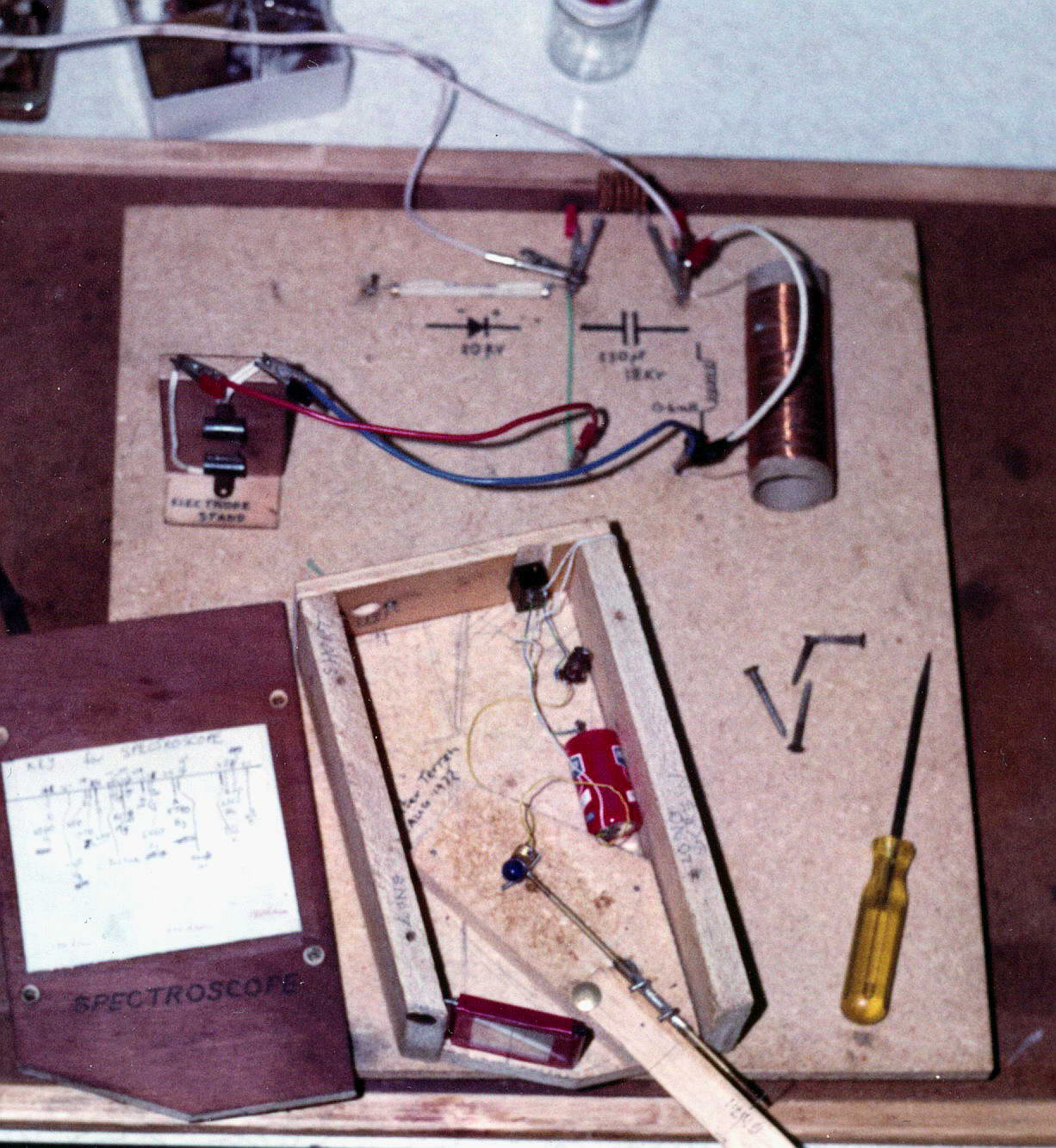
My first TC was ‘discovered’ while running a spark spectroscopy project (above) in high school 40 years ago. This used about 10 kV from multivibrator excited dual ignition coils through 10 turns of an air cored radio coil to quench it. The other end of the 130 turn coil developed a corona visible in the dark spectroscopy room. It was a truly amazing sight.
The spark spectroscopy setup in use (but not in operation as a Tesla coil). The original high voltage setup with the spectroscope shown open and the HV diode, air cored coil and cap made of 6 x 0.0033 uF 3 kV ceramics in series to give 550 pF. The spark gap with clips to hold the metal being examined spectroscopically is also shown.
Related pages
Try something else
Tesladownunder FAQ (frequently asked questions)
External links
Emission spectra applet
Spark and arc atomic emission spectroscopy
Photo Date: May 10, 2010; May 16, 2010
